|
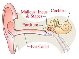
In The Inner Ear,The Cochlea Curls Around Like A Snail Shell..........
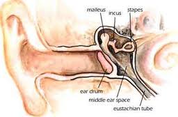
The Inner EarThe inner ear is not a removable structure that could stand alone, like the ossicular chain. It is a series of tunnels and cavities hollowed out of the petrous portion of temporal bone. These tunnels are filled with two separate and continuous fluids from one portion to another. The outer fluid is always perilymph. The inner fluid, which is separated by a membrane, is endolymph fluid. Although it is a continuous cavity, it is discussed as if it were three separate and distinct parts, semi-circular canals, vestibule and cochlea.
There are three semi-circular canals, Superior, Posterior and Lateral, oriented at 90 degrees to one another in the inner ear. The outer portion of those fluid filled ducts contains perilymph fluid, which protects the membranous endolymph fluid. These canals detect rotational movement in any direction for balance and equilibrium. The canals serve no known hearing function. Balance problems and dizziness or vertigo can develop, along with
hearing loss
as the fluids are continuous throughout the inner ear.
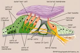
The CochleaWithin the inner ear, the cochlea coils around like a snail shell in 2-2.75 turns. The inside of this bony labyrinth sections off, but is not completely divided, into three parts by the (bony) osseous spiral lamina on the inside wall, and the spiral ligament on the outside wall. These sections consist of three tapering tubes (scalae) in layers, one on top of the other. The two outer tubes, containing perilymph fluid, are the scala vestibule on the top, and the scala tympani on the bottom. The middle smaller, triangular shaped tube is called the scala media. The auditory nerve endings are in the scala media, also called the cochlea duct, or canal of the cochlea, or membranous labyrinth. The Organ Of CortiIt consists of columns of inner and outer rods with hair cells (cilia) at the top of the rods. Supporting cells hold these rods and hair cells in place. Although illustrations show only a few hairs in the cells, they are more like a hairbrush, with tufts of 40 hairs at the base of the cochlea, increasing to tufts of 100 at the apical end. There is one row of inner rods and three to five rows of outer rods depending on the location of the cochlea. The tectorial membrane, a gelatinous structure, attaches to the supporting cells at the inner side only, and is suspended over the hair cells. Tufts of the tops of the hair cells are embedded in the underside of the tectorial membrane. Any vibration through the surrounding fluid, or movement of the tectorial membrane, affects the hair cells. The fluid vibrations cause the tectorial membrane to undulate. A shearing action takes place on the hair cells. The bases of the hair cells contain nerve endings, converting this shearing action into nerve impulses. At this point, the nerve impulses become electrical energy. The cochlea is often compared to a piano. The piano has only eighty-eight keys, while the cochlea in effect, has thousands. The piano covers tones of a little more than seven octaves. The cochlea covers almost one octave lower than the piano and slightly more than two octaves higher. The base of the cochlea, nearest to the oval window, responds to high frequencies. The part of the cochlea farthest away, at the apex, responds to low frequencies. Intermediate frequencies occur at intervals between the base and the apex.
Hair CellsHair cells are arranged in well defined rows, with about 3500 inner hair cells and 12,000 outer hair cells. The outer hair cell are believed to be 35db more sensitive than the inner hair cells, are damaged by loud sounds or ototoxic drugs. It pays to protect your hearing.
The base of the hair cells contains nerve endings, which, when triggered or excited become nerve impulses. The loudness of sound appears to relate to the number of nerve fibers that are stimulated and to the rate of stimulation. The hair cells play a vital role within the inner ear. RetrocochlearThe auditory nerve (v111 Cranial Nerve) consists of 30,000 neural fibers (neurons) with 95% of these fibers connecting to the inner hair cells, and 5% to the outer hair cells. The neurons are afferent fibers. They transmit from the cochlea to the brain. Efferent fibers travel from the brain to the cochlea. The function of the efferent fibers is not known. By now, it is fairly clear that the ear does not hear and understand speech. The ear transmits frequencies and intensities to the brain. The brain translates these frequencies and intensities in something meaningful,
speech,
noise,
|